
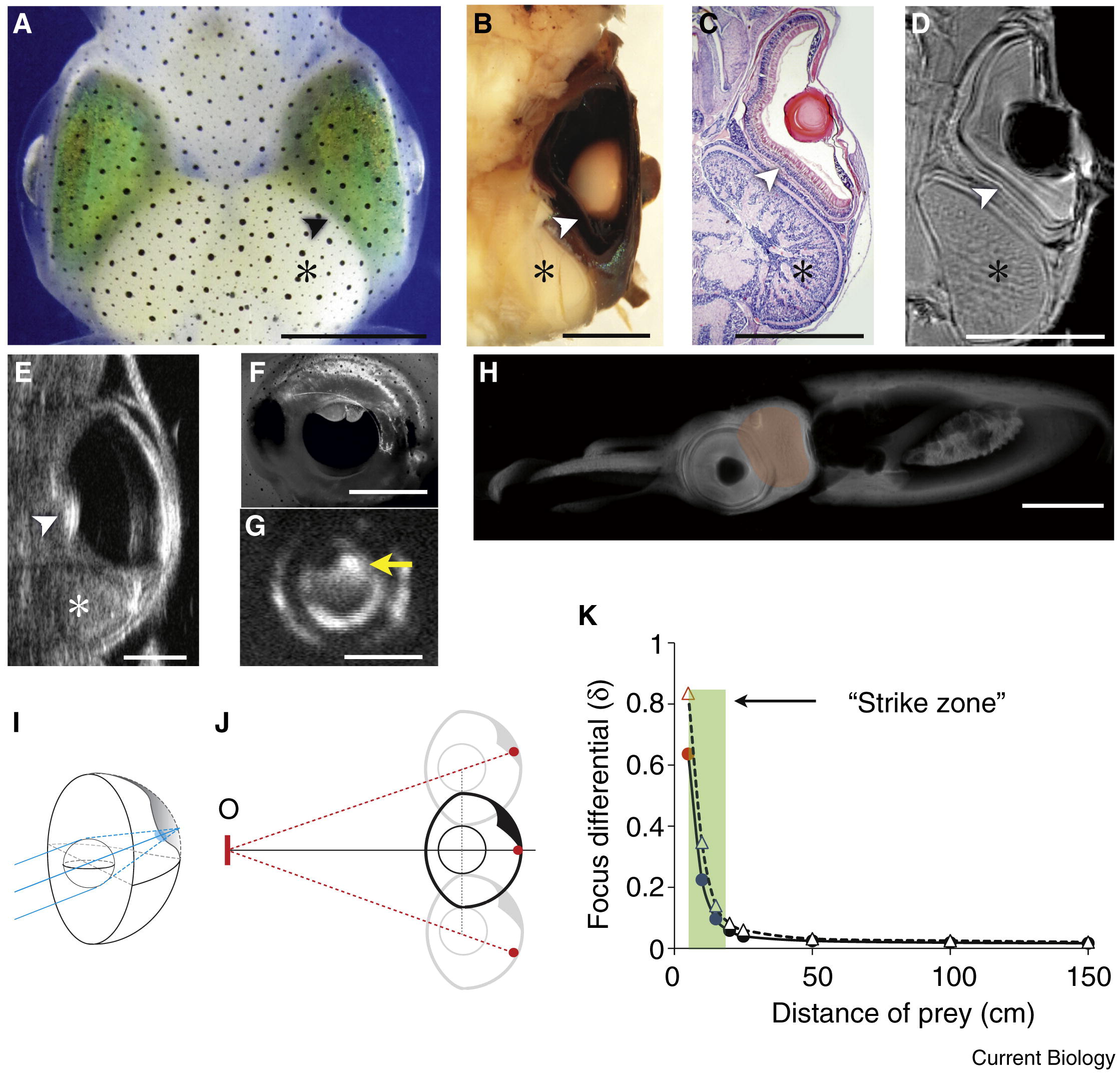
The non-hemispherical eye resulting in a new form of range-finding in Sepioteuthis lessoniana.
(A–H) The retinal deformation of squid permanently appears using a combination of techniques. (A) The live specimen. (B) Dissection of the rapidly fixed eye and brain using perfusion. (C) Histological cryosection of an unfixed, fresh specimen (12 μm thickness). (D) High resolution contrast-enhanced MRI of a lightly preserved specimen. (E) High resolution ultrasound imaging of an anesthetized specimen. The retinal bump (RB) (arrowheads) and optic lobe (OPL) (stars) are indicated. (F) The dilated pupil under red illumination. (G) Retinoscopic imaging of free swimming squid. The bright reflection on the upper area indicates hyperopic defocus (arrow). (H) MRI sagittal section. The shaded area (pink) shows that the OPL intrudes into the back of the retina at the dorso-temporal region, causing the frontal and downward (approximately 35°) view to be defocused. Scale bars: 5 mm. (I–K) Object distance estimation using retinal bump and head bobbing behavior. (I) Schematic drawing of the anatomical model of the deformation of the eye and the resulting defocus on the retinal bump (blue circles). (J) Dynamic eye positioning places the object’s image (O) in and out of the retinal bump, resulting in changes of the focal quality (see Supplemental Information for further details). (K) The curves show focus differentials of a particular object over distance for both a constricted pupil (diffraction-limited condition; solid line) and a dilated pupil (diffraction-free condition; dashed line) (see Supplemental information for further details). In both conditions, the focus differential remains low for distant objects until 15 cm (blue circle), the point just within tentacle range (specimen mantle length, ML): 5 cm, tentacle projecting up to 2 MLs).

